- Ultrasound for confirmation pregnancy
- Ultrasound for excluding Down syndrome and measuring the nuchal translucency thickness of the fetus:-
- Determination twins pregnancy :-
- Ultrasound for exclusion any abnormalities in all fetal parts:-
- Diagnosis of recurrent pregnancy loss:-
- Fetal Echocardiography :-
- Ultrasound for measuring the cervical length :-
- Ultrasound for determination fetal growth and fetal position before delivery :-
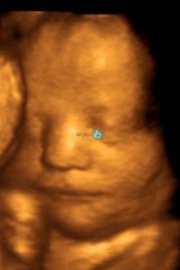
- Ultrasound 3D – 4D:-
The goal of this ultrasound:-
It is to confirm the state of pregnancy and the number of fetuses present, and to ensure the normal growth of the fetus inside the womb and not outside the womb, and shows the state of twins.
This sonar is very important and necessary for women who suffer from pain or bleeding during the first months of pregnancy and for women who suffer from repeated miscarriages and ectopic pregnancy.
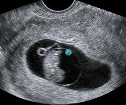
Picture No. 1 shows the presence of the beginning of the formation of a fetus inside the pregnancy sac inside the womb
Picture No. 2 shows an empty pregnancy sac with no fetus

Dr. Wael Al-Banna makes this ultrasound from the 11th to the 14th week of pregnancy. Through the abdomen, but in some cases, the vaginal ultrasound is used.
The goal of this ultrasound :-
For determination of gestational age accurately.
This is for women who do not remember their last period, or who suffer from irregularity in the cycle or have a pregnancy while breast feeding, or a pregnancy occurred after stopping the contraception pill.
In this ultrasound, you measure the size of the fetus accurately and calculate the gestational age and expected date of birth.
To take advantage of this ultrasound:
Determining and evaluating the probability of the fetus being infected with Down syndrome or fetal abnormalities.
Each woman will give the ratios of the probabilities that her fetus will have Down syndrome or various genetic syndromes taking into consideration:
1- The age of the woman.
2-Measuring hormones in the blood of a pregnant woman.
3-The thickness of the fetus’s nuchal translucency
4-Fetal echocardiography.
5-Blood flow rates in the heart.
6-doppler for ductus venosus.
Parents will receive full instructions on the condition of their fetus, and if the fetus is dangerous, it needs to perform a sample drawn from the amniotic fluid or do a fetal DNA analysis, ablood sample that is from the mother and genetically analyzed, and shows whether the fetus has a genetic syndrome or not
And the advantage of this analysis from taking a sample of amniotic fluid. It is to reduce the incidence of miscarriage that the mother will be exposed to when taking a sample of amniotic fluid, but the cost of this analysis is much higher.
2% of women who became pregnant naturally could carry twins.
10% of women who used ICSI or normal induction activate twins.
This ultrasound can at this time know whether or not the fetus is developing normally. Do they share the same placenta or not?
And this ultrasound is done in the 5th week of pregnancy.
If there is any abnormality in ultrasound finding, patient must follow up sooner.

This detailed ultrasound is performed by Dr. Wael on week 20-24 of pregnancy.
During this ultrasound we can show:
1- Placental site
2- Amniotic fluid index
3- Fetal weight and fetal growth
4- Detailed examination for brain ,heart , vertebral column, stomach and extremities of the fetus.
If a congenital anomaly is identified that necessitates fetal surgery in the womb , the doctor himself performs this surgery.
Unfortunately, it was found worldwide that 2% of women who do sonar work the fetus has died since previous weeks and without any warning.
At this time, the couples receive instructions and analyzes are made to find out the cause of death.
During ultrasoun of the Week (11-13), ultrasound of the Week (20-24) and ultrasound of the Week (30-34)
We routinely make ultrasound on the heart of the fetus and the blood vessels in the heart of the fetus.
Who are the women who should make ultrasound on the hearts of their fetus?
1-Women with family history of congenital heart diseases.
2-Women who suffer from diabetes with pregnancy.
3-Women who take epilepsy and psychological drugs with pregnancy.
4-Fetuses found in the first ultrasound of pregnancy having an increase in the rate of nuchal translucency in the fetus.

Dr. Wael makes a vaginal ultrasound to measure the length of the cervix
This ultrasound is essential and required for all women who have suffered from premature birth or are pregnant with twins or who have congenital anomalies in the womb before (the bicornuate uterus – or women who have undergone cervical operations).
This ultrasound is done by Dr. Wael to make sure:
1- Fetal growth in the required size at the end of pregnancy
2- Check Fetal movement and 4-D video recording.
3- Determine the placental site before birth.
4- Measuring the amniotic fluid index around the fetus.
5- Determining the blood flow rate ( umbilical doppler) to the fetus and to the organs of the fetus.
This ultrasound examination is usually performed over the 32 weeks of pregnancy.
Dr. Wael is recommended for women who have complications of pregnancy such as growth retardation, diabetes, and for women who have any abnormal data during the current pregnancy.
This ultrasound aims to determine the growth and health of the fetus before:
1- Measurement of fetal head size, femur and abdomen, and calculation of fetal weight
2- Examination of fetal movements
3- Evaluation of placental site and appearance
4- Measuring the amount of amniotic fluid
5- Evaluation of blood flow to the placenta and fetus by ultrasound color Doppler